RP is the world’s most prevalent inherited retinal disease and a leading cause of visual disability and blindness, often beginning in childhood or adolescence. Resulting from diverse inherited genetic mutations, RP causes progressive, irreversible vision loss, profoundly impacting a patient’s autonomy and quality of life.


More than 1.5 million people affected worldwide
1 in 4,000 people affected
Over 100 causative genes (including nonsyndromic forms)
Symptoms often begin in childhood or adolescence
Progressive vision loss over years to decades
No curative treatment

Patient Experience1-6
Gradual, often subtle onset
RP typically begins quietly. Many patients first notice trouble seeing in dim light or at night, while daytime vision seems normal. Early on, reading and central vision are usually preserved, making the condition easy to miss or dismiss as poor night vision.
From night blindness to tunnel vision
As RP progresses, the loss of rod photoreceptors leads to shrinking peripheral vision. People may bump into objects at the side, struggle in unfamiliar environments or lose confidence behind the wheel, especially at night. Over years or decades, the visual field can contract into “tunnel vision,” and later stages may compromise central vision as cone cells are affected.
Living with RP vision loss
RP often unfolds during school years or early adulthood, at a formative time for personal and professional life. Its progression can limit autonomy and affect career choices and employment options. This disruption subjects many RP patients to depression, anxiety, and social isolation, and can weigh heavily on their families.
Understanding the Disease Mechanism1, 7, 8
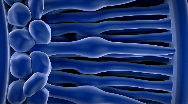
The genetic origin
RP is caused by mutations across more than 100 distinct genes (including syndromic forms), spanning autosomal dominant, recessive and X-linked inheritance patterns. These mutations disrupt biological pathways essential to photoreceptors or to the retinal pigment epithelium that supports them. Photoreceptors are the retinal cells responsible for detecting light and converting it into electrical signals for the brain.
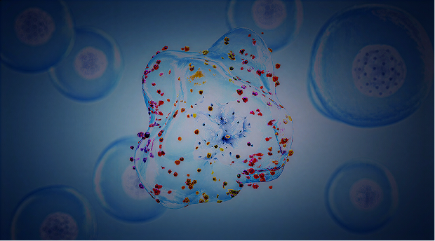
Oxidative stress and cellular damage
In most forms of RP, rod photoreceptors, which manage night and peripheral vision, degenerate first. This initial cell loss triggers metabolic imbalance, oxidative stress and loss of supportive factors across the retina, which subsequently damages the neighboring cone photoreceptors that are involved in detailed and color vision.
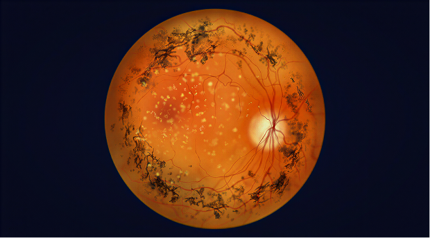
Vision loss
As photoreceptors degenerate, fewer light signals are relayed through the retina to the optic nerve. The brain receives increasingly fragmented visual information, translating this cellular decay into patchy blind spots, progressive tunnel vision and, in many patients, eventually severe vision loss or legal blindness.
The RP Patient Journey
Historical Perspective
Described in the 19th century from its characteristic pigment deposits and narrowed retinal vessels, RP was long understood only as a clinical pattern. Advances in clinical research transformed RP into a prototype inherited retinal dystrophy. Today, it is recognized as the most common inherited retinal disease and a key model for understanding genetic blindness.
Current Treatment Landscape
There is currently no cure that halts or reverses RP for most patients. Supportive care, such as low-vision aids, orientation and mobility training, and environmental adaptations, remains central to preserving independence. The first gene therapy, voretigene neparvovec, is approved for a small subset of patients with biallelic RPE65 variants, marking an important milestone but addressing only a fraction of the RP community. Numerous clinical trials in different therapies are underway to meet this high unmet medical need.
Diagnosis & Patient Care
Diagnosis typically begins with a comprehensive eye examination, followed by genetic testing to pinpoint the causative variant when possible. Suspected cases should be referred to a retina specialist or inherited retinal disease center for confirmation and counseling. Early recognition allows patients to access low-vision services, genetic counseling, and participation in clinical research, helping them plan for the future and benefit from emerging therapies.
Get in touch with our team to learn more.
REFERENCES:
-
Verbakel SK, van Huet RAC, Boon CJF, den Hollander AI, Collin RWJ, Klaver CCW, et al. Non-syndromic retinitis pigmentosa. Prog Retin Eye Res. 2018;66:157-86. doi:10.1016/j.preteyeres.2018.03.005
-
Pniakowska Z, Dzieża N, Kustosik N, Przybylak A, Jurowski P. Genetic therapies for retinitis pigmentosa: current breakthroughs and future directions. J Clin Med. 2025;14(16):5661. doi:10.3390/jcm14165661
-
Colombo L, Baldesi J, Martella S, Quisisana C, Antico A, Mapelli L, et al. Managing retinitis pigmentosa: a literature review of current non-surgical approaches. J Clin Med. 2025;14(2):330. doi:10.3390/jcm14020330
-
Heath Jeffery RC, Lo J, Thompson JA, Lamey TM, McLaren TL, DeRoach JN, et al. Driving with retinitis pigmentosa. Ophthalmic Genet. 2023;44(4):352-60. doi:10.1080/13816810.2023.2196338
-
Abu Serhan H, Ahmed A, Sbei O, Kojan W, Chaudhry M, Khalid M, et al. Mental health outcomes in patients with inherited retinal diseases: a systematic review and meta-analysis. Int J Retina Vitreous. 2026;12(1):45. doi:10.1186/s40942-026-00820-7
-
Garip G, Kamal A. Systematic review and meta-synthesis of coping with retinitis pigmentosa: implications for improving quality of life. BMC Ophthalmol. 2019;19(1):181. doi:10.1186/s12886-019-1169-z
-
Demirkol A, Kendir Uguz F, Cavus NM, Demirkol I, Tsang SH. Clinical characteristics and genetic factors in retinitis pigmentosa: a retrospective analysis of a Turkish patient cohort. Med Sci (Basel). 2026;14(1):24. doi:10.3390/medsci14010024
-
Campochiaro PA, Mir TA. The mechanism of cone cell death in retinitis pigmentosa. Prog Retin Eye Res. 2018;62:24-37. doi:10.1016/j.preteyeres.2017.08.004
